This post is third in a series highlighting a selection of The Mark Foundation for Cancer Research’s scientific partners who have rapidly mobilized resources to confront the great challenges posed by COVID-19 (you can see the earlier posts here and here).

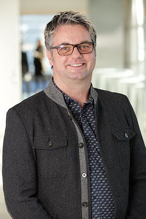
Recently, I caught up with our Scientific Advisory Committee member Jeroen Roose, PhD, Principal Investigator and Vice Chair of Anatomy at the University of California, San Francisco, to learn about his lab’s efforts to characterize the effects of SARS-CoV-2 on lung cells and understand the response of the immune system to infection.
Jeroen’s research focuses on understanding cell signaling in autoimmune diseases and cancer. In particular, his lab studies the role of Ras-kinase signaling pathways in cell fate and disease. His lab consists of immunologists, cancer biologists, biochemists, and cell biologists who collaborate in teams. Recently, many team-projects are capitalizing on organoids, 3-dimensional mini-organs grown from human or animal cells in a cell culture dish. To study cancer, Jeroen’s team generates organoids with cells from patients undergoing tumor resection. Organoids are useful for understanding biology because they adopt some of the architecture of the tissues from which they originate and, therefore, more faithfully represent the physiology and pathology of those tissues. Organoids are also genetically stable when cultured in the right conditions, maintaining stem cell functions but also patient-specific properties. The experts in the lab can create organoids from cancers such as colorectal, pancreatic, breast, and lung. Wisely, they are freezing organoid material to assemble a biobank of genetically and pathologically diverse models that can be exploited for multiple purposes. The lab also obtains blood samples from the patients whose cells are procured, allowing them to probe deeper into associated aspects of patient biology and disease.
To source the cells for lung organoids, Jeroen collaborates with UCSF colleague Johannes Kratz, MD, who specializes in minimally invasive robotic surgery. Typically, when a tumor is surgically removed, adjacent unaffected tissue is also obtained. Because the lab gets both tumor and normal tissue, they can grow both types of organoids as so-called genetic twins, which has been invaluable in uncovering the biology that differentiates tumors from non-cancerous lung tissue. As the pandemic surged and Jeroen faced the soul-crushing mandate to shut down most of this work, he pivoted quickly to deploy the normal lung organoids to understand SARS-CoV-2 viral infection and look for potential treatments.
Lung cells are a type of epithelial cell, cells that line the surfaces and cavities of body tissues and organs. Using the organoids, Jeroen and colleagues are uncovering the pathways and cell biology triggered in epithelial cells upon SARS-CoV-2 infection and aim to study why people react differently to the virus. The beauty is that lung organoids from different individuals are all unique, creating a museum of mini-lungs in a dish. They will now infect these with different SARS-CoV-2 strains from patients in San Francisco and from patients in Marseilles, France provided by collaborators Jean-Pierre Gorvel, PhD, DRCE, CNRS at Centre d’Immunologie de Marseille-Luminy (CIML) and Jean-Louise Mege, MD, PhD at Institut Hospitalo-Universitaire (IHU). These strains are genetically different, which could be meaningful for COVID-19. The San Francisco strains resemble strains identified in Wuhan, China whereas the strains from Marseilles are more similar to those isolated from patients in New York City. Using H1N1 influenza virus as a comparator, the team is looking for contrasts and commonalities in the responses of epithelial cells to infection. For example, when infected with virus, epithelial cells secrete factors that recruit immune cells to help fight the infection. It is possible that these responses are stronger or weaker depending on the strain of the virus and/or the genetic or cellular makeup of the infected lung organoids.
The team will also explore what happens as cells within organoids are infected, die, and then are replaced by proliferating stem cells that differentiate into new cells with specialized functions, reflected in interesting biological names: basal, club, goblet, and ciliated cells. They specifically want to know whether the newly generated cells will also get infected and if they don’t, how they have altered cellular processes or signaling pathways to garner this protection. These insights could lead to paths for new or existing therapies that mimic these protective effects within lung cells.
At a later point the study involves co-culturing immune cells from the same patient’s blood with the infected organoids. This approach could allow the team to identify and expand immune cells that are reactive to virus-infected cells. These immune cells will be a rich source of information that could shed light on the biology of effective versus ineffective immune responses. The team will focus on T cells as they are experts in the signaling pathways that influence T-cell biology. In this respect, their COVID-19 research has many similarities with their cancer research.
UCSF has amazing infrastructure for these studies, so-called “CoLabs”, which will allow Jeroen and colleagues to carry out high-resolution imaging of the co-cultures, analyze changes in gene expression in the tissues (and even the individual cells involved), work under BSL3 conditions (the highest safety level for work with pathogens), as well as deploy a sophisticated platform called CyTOF, which can provide a very detailed analysis of the immune cells’ characteristics and how those change for better or worse during the lifecycle of the infection. These insights could lead to paths for new or existing therapies that could tune the immune system to productively fight viral infection.
The excitement about this work was very evident when I spoke with Jeroen. It’s important to point out that this pivot to COVID-19 is not replacing his cancer research. While, unfortunately, cancer research has slowed down in many cases across the globe during the pandemic, the knowledge gained through COVID-19 work will advance knowledge in the realm of cancer research as well, as it will help us better understand immune system responses to diseased tissue. Some of those learnings will undoubtedly be relevant in the context of cancer. In addition, Jeroen’s lab will take advantage of the genetic twin cancer-derived organoids in their biobank to study what happens to tumor tissue in the lung that most certainly will be vulnerable to viral infection and extrapolate to what that might mean for cancer patients exposed to SARS-CoV-2.
Finally, pivoting one’s lab cannot be successful if there are limited resources to fund the work. Jeroen is especially grateful to the NIH for the foresight to allow researchers to draw from and supplement their existing grants so they can move nimbly into this space. However, this funding truly represents only a seed of what is needed. As results from labs across the nation that have likewise pivoted start to emerge, it will be up to biopharma companies to accept the baton and keep running as fast as possible.


